Spontaneous Pneumothorax:
In infected peoples’ lungs, there are these blebs from the tissue that are formations of little sacs/ pockets of air, and these people could have 1 to more than 30 of these ugly balls of airs. These sacs of air can explode, causing leak of air into the pleural space – which is the space between your lung and your pleura – the outer layer of the lung. When the air leak out to the pleural space, the air will increase and force the lung to compress into smaller size, and it could even compress as small as a quarter of your original lung size.

Tension pneumothorax usually happens when trauma is applied to the lungs, puncturing the lung. The same thing happens to the lung as the spontaneous pneumothorax, the lung got punctured and the air go to the pleural space, compressing the lung.

Hemothorax:
A hemothorax usually happens in blunt force trauma or penetration. A blood vessel will rupture, an intercostal vessel or an internal mammary artery. Hemorrhage comes in different ranges when the vessels rupture and the massive hemorrhage could even cause a shock. Blood then fills up the pleural space, pushing the lungs, and forcing it to compress.
Symptoms:
Symptoms:
Pneumothorax:
-Short of Breath
-Fatigue
-Bluish Skin
-Low Blood Pressure
-Chest Pain
-Tight Chest
-Swollen Abdomen

Hemothorax:
-Rapid Breathing
-Breathing Difficulties
-Bluish Skin
-Chest Pain
-Low Oxygen Levels

Diagnosis needed:
Tension Pneumothorax: Clinical evaluation (Pearls and pitfalls)
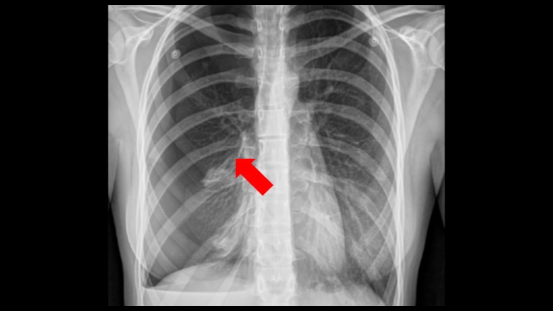
Spontaneous Pneumothorax: Chest X-Ray/CT(computed tomography)
Hemothorax: Chest X-Ray
Causes:
-Rib Fractures (T)
-Chronic Obstructive Pulmonary Disease (S)
-Cystic Fibrosis (S)
-Pneumonia (S)
-Ruptured Air Blisters (S)
-Blunt Force Trauma (H)
*T=Tension, S=Spontaneous, H=Hemothorax)
Surgery Cure:
1. Wear protective clothing
2. Clean skin with solution
3. Apply Anaesthesia to the patient (Anaesthesia=drug that makes the patient unconsious)
4. Mark the insicion spot (4th or 5th intercostal space) with marker
5. Cut with No. 10 Blade
6. Put chest tube (which is connected to chamber) into the hole
7. Let it suck the fluid out
8. Remove tube after less than 100 ml fluid/air flow into chamber
9. Stitch the wound up.
Although surgery can cure it, there are still risks
Risks of Surgery:
- -Lung damage
- -Other organs damage
- -Bleeding/ infection to chest tube wound
- -Wrong placement of chest tube
No comments:
Post a Comment